Oral and Maxillofacial Surgery Technology

Our office is fully equipped throughout with cutting edge technology. This technology is maintained and upgraded regularly. We are confident that you will appreciate our commitment to modern technology and see how it directly improves treatment and care of our patients.
Use of Electronic Medical Records enables our practice to quickly and accurately access patient information. This helps to ensure patient confidentiality as well as reduce the need for paper. Using a digital format allows for quick access to patient information when needed for insurance records while providing a secure filing system.
An intraoral camera combines the latest video technologies with oral surgical care. Digital photography allows both the doctor and our patients to immediately view detailed images of their mouth or teeth in real time. With an intraoral digital photography patients are able to better understand and appreciate their condition and the status of their oral health.
Our state of the art handpieces and saws are the most efficient and powerful handpieces. They provide less vibration and noise than traditional dental drills.
These handpieces are specialized sophisticated ultrasonic saws used specifically for cutting bone atraumatically. These handpieces gently and meticulously cut bone and enable the doctors to perform improved services never before performed in an office environment.
Our 3 offices are located in Hudson and Bergen counties and these offices are completely technologicaly integrated. All of our patient records are available in any of our locations at all times. The offices are equipped throughout with digital flat screen monitors. With these monitors our patients can easily view their digitalized imaging studies as well as any digitalized photography when speaking with the doctor. Clear viewing of clinical findings provides more appreciation and a better understanding of their oral health.
This section will provide you with a short description of the different types of imaging instrumentation that our office provides. Over the years our offices have always been equipped with the most up to date imaging equipment available to provide our patients with the most efficient, dependable images with the least amount of radiation exposure. Today the imaging studies available have evolved dramatically as the type and quality of the images that are available is quite impressive. We have maintained our “state of the art” reputation and recently upgraded all units throughout our practice. In addition our offices now, are all totally linked electronically, so all radiographic studies performed at any location are readily available at the time of your office visit.
Currently all of our offices are 100% digitalized and we are equipped to perform the following film studies within the confines of our offices:
- Periapical radiograph: This x-ray is the conventional dental radiograph (small dental radiographs) and is used in our office on occasion.
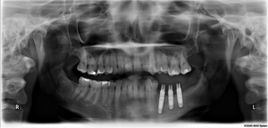
- Orthopantomogram (Panorex): Panoramic imaging remains the standard of care for many types of dental procedures. In todays medical world the panorex is without question the most comprehensive preliminary screening imaging study available. It is essential for almost any type of oral surgical procedure. This study provides us with a complete over view of the lower 1/3 of the facial skeleton provides an enormous amount of diagnostic information with minimum radiation exposure. It remains the back bone of oral surgery and the least invasive (with respect to radiation exposure and patient acceptance). Panoramic units are in place at all three office locations and are of the highest quality, emitting the least amount of radiation while providing us with the necessary diagnostic information.
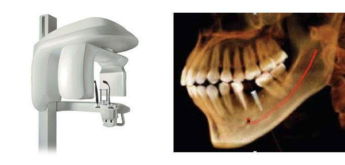
- Our most recent advancement is our Cone Beam Computerized Tomographic (CBCT) machine located in the Hackensack office. This unit is a Kodak 9300CT and can provide a fully digitized 3-D study of the entire facial skeleton. The Kodak 9300CT is the most sophisticated cone beam unit available on todays market and while these units are becoming popular, there are a number of distinguishing features about our unit which make it the most sophisticated unit available at this time:
- The lowest level of radiation emissions per study.
- The 9300CT enables us to isolate our study, if necessary, to a small area of interest rather than “shoot the whole face”. From our point of view this feature is critical with respect to our patients overall health and welfare as we are able to effectively minimize radiation exposure to our patients and scan small isolated areas throughout the facial skeleton when necessary.
This new addition to our diagnostic armamentarium provides us with cutting edge technology that enables us to provide a clear and more definitive diagnosis (especially with respect to implant restoration and facial reconstruction) and more accurately anticipate potential surgical sequel.
The Carestream 9300CT is the most sophisticated cone beam unit available on today’s market and while these units are becoming popular, there are a number of distinguishing features about our unit which make it the most sophisticated unit available at this time:
- The lowest level of radiation emissions per study. Your health and safety is our greatest concern.
- The 9300CT enables us to isolate our study, if necessary, to a small area of interest rather than “shoot the whole face”. From our point of view this feature is critical with respect to our patients overall health and welfare as, once again we are able to effectively minimize radiation exposure to our patients and scan small isolated areas throughout the facial skeleton when necessary
- High resolution images that allow us to see your teeth and facial skeleton with unprecedented detail
- Comfortable positioning: the system’s open design makes exams more comfortable, with both seated and standing options to accommodate patients of all sizes and the unit is wheelchair accessible
- Improved care: we can perform a wider range of diagnoses with even greater accuracy
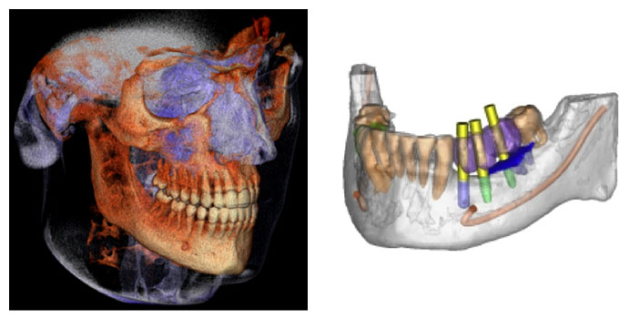
The new addition of this versatile 3D system to our diagnostic armamentarium provides us with cutting edge technology. You will see this “cutting edge technology” is evident throughout all aspects of our practice and it allows us to meet the full range of our patients imaging needs. In addition to the 3D images, this 3-dimensional system will also deliver crystal clear 2D panoramic radiographs (conventional x-rays), which still form the diagnostic foundation of most cases. These images enable us to provide a clear and more definitive diagnosis (especially with respect to implant restoration and facial reconstruction) and more accurately anticipate potential surgical sequela. Because many cases require additional anatomical detail, the system’s multiple 3D imaging capabilities support a wide range of clinical applications with focused-field, single and dual jaw views. TMJ, sinus and maxillofacial views are easily obtainable. This state-of-the-art technology will help us diagnose with an enhanced level of accuracy and provide treatment with unprecedented confidence. Whether we are analyzing a complex tooth impaction, planning a multi-site implant case, or visualizing TMJ dysfunction, the precise, crystal-clear 3D and panoramic images will help us to formulate a specific treatment plan that will address your needs and answer any questions that you may have about your care and treatment options.
This new addition to our diagnostic armamentarium provides us with cutting edge technology that enables us to provide a clear and more definitive diagnosis (especially with respect to implant restoration and facial reconstruction) and more accurately anticipate potential surgical sequel.


